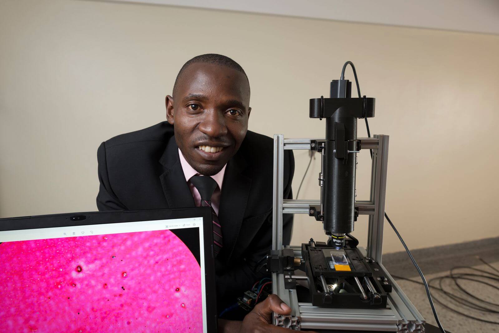
Conventionally, pap-smear images are analysed manually, which is time consuming, error-prone and has to be done by a trained cytopathologist. Cervical cancer risk factor analysis is also not incorporated into this process, and the digital microscopes required are expensive, which means they are few and far between in low-income countries. Low cost microscopes are used, but then slides can’t be digitally stored for analysis.
Dr Wasswa, the head of Mbarara University’s biomedical engineering department at only 30 years old, developed a digital microscope slide scanner to quickly scan high-resolution cervical cell images from pap-smears. The youngest engineer to complete his doctorate at his university, Wasswa 3D-prints the parts for the device, which costs nearly a quarter of the price of commercial microscopes.
Not satisfied with merely speeding up the scanning of cells, Wasswa has also created an analysis tool for diagnosis and classification of the images. His system, which has been published in peer-reviewed academic journals, has had between 90 and 100% accuracy during testing.
Building on the knowledge base of doctors and experts, Wasswa went further to design software which automatically assesses the likelihood of a patient contracting cervical cancer given their risk factors, and a separate system for managing and archiving patients’ records using artificial intelligence.
The PapsAI system is currently being tested at a local hospital, and Wasswa hopes it will bridge the healthcare gap by providing relevant solutions at an affordable price.


